By YAN Fusheng (Staff Reporter)
Tracing proliferative cells in vivo may inform richly to many biological or pathological events. However, current approaches are yet mostly insufficient to get a clear picture of cell proliferation either at the scale of whole cell population or in a cell-specific manner, which could inevitably stain the conclusion. To this end, CAS scientists developed a powerful method of proliferation tracing, dubbed ProTracer. Highlighting its capabilities, they settled a long-standing debate – which zone of liver is the main source of repopulating hepatocytes during homeostasis.

Enabled by proliferation tracer (ProTracer), a new genetic proliferation lineage tracing method, researchers settled a long-lasting debate – cells from which zone of the liver lobule are the major contributor to hepatocyte proliferation during liver homeostasis. As found in mice liver, the newly generated cells (marked as red spots), or the hepatic stem cells, are mostly located in the midlobular zone. (Image by ZHOU’s Lab)
Cell proliferation, one cell divides into two cells, is the basis for a fertilized egg to grow into a fetus. It is also the reason why we can heal from a cut, or a salamander can regrow its tail. When cell proliferation goes crazy, it is most likely that a cancer is being hatched.
The ability to monitor cell proliferation is therefore essential for a myriad of studies in developmental biology, regenerative medicine, and oncology. Therefore, scientists have put many efforts in developing different approaches for proliferation tracing. However, the limitations of current approaches for measuring cell proliferation in vivo have left many fundamental questions in numerous life science fields insufficiently addressed. The inherent bias of
Applying these approaches to probe cell proliferation in vivo is, most of times, like “the blind men feeling an elephant”. The inherent bias could easily lead to conflicting conclusions, as illustrated by the highly controversial studies around cell sources for hepatocyte generation.
To address these unsettled questions, a team of CAS scientists, led by Dr. ZHOU Bin from the CAS Shanghai Institute of Biochemistry and Cell Biology, Center for Excellence in Molecular Cell Science, reported a new strategy to inducibly and permanently label specific proliferating cells with a fluorophore, dubbed ProTracer.
ProTracer permits the temporally continuous genetic recording of in vivo cell proliferation in diverse cell lineages over time in multiple organs and tissues. Like a video recorder, ProTracer, once turned on, can record cell proliferation continuously in any defined time window through an animal’s life span by lighting up proliferating cells with a permanent expression of a fluorescent protein. This unique feature makes ProTracer extremely useful in detecting stem cells with low proliferative capacity, which is quite challenging for current approaches.
ProTracer can also be primed to exclusively trace proliferating cells in a cell-specific manner, which avoids interference from proliferation signals of other types of cells and thereby brings the footage into sharp focus. The additional bonus of ProTracer is that it is noninvasive and free of toxicity, hence friendly with experiment animals.
Highlighting its capabilities, the researchers tuned ProTracer to specifically record proliferation in hepatocytes by using a hepatocyte-specific promoter. By doing so, they settled a long-lasting debate – which zone of the liver lobule, the repeating architectural element of liver, contributes the most to repopulate hepatocytes during liver homeostasis and repair. By ProTracer-based continuous recordings over several weeks to months, a population of hepatocytes located in the midlobular zone (i.e., zone 2) is identified with more proliferative capacity during liver homeostasis in mice, whereas zone 1 and zone 3 hepatocytes predominantly respond to injury and repopulate the liver after injury to zones 3 and 1, respectively. These newly-gained insights may brew new therapies that aim to activate specific cellular compartments responsible for regeneration.
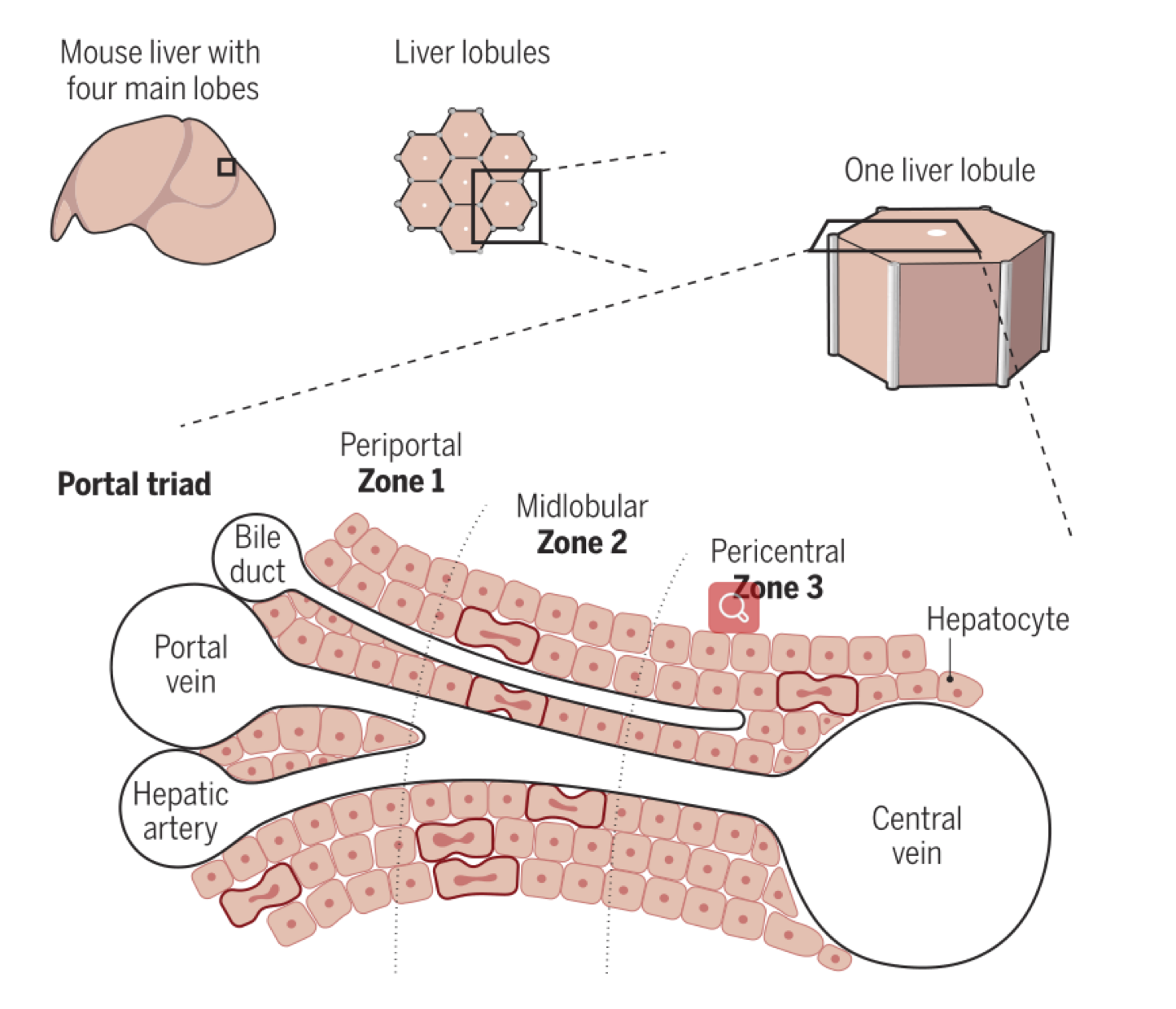
Liver architecture is defined by repeating units, called liver lobules, which have a hexagonal structure with a central vein and a portal triad (portal vein, hepatic artery, and bile duct) at each corner, separated by hepatocytes. Hepatocytes exhibit specialized functions from the portal to central vein, spanning three zones from periportal (zone 1) to midlobular (zone 2) to pericentral (zone 3). (Image by E. R. Andersson)
“The ProTracer mouse model opens up the possibility of tracing cell proliferation from adult stages over longer time scales (several months) in a continuous manner, enabling the identification of slowly cycling cells and rare stem cells in various organs. This may unveil previously unknown stem cell populations,” commented Emma R. Andersson, a scholar from Karolinska Institutet, who is not involved in this study, in the same issue of Science.
Enabled by this new recording tool on proliferating cells, many more interesting cellular behaviors once veiled may become discernable to good eyes, which would then intrigue scientists to unravel their underlying mechanisms at molecular levels.
References
L. He, W. Pu, X. Liu, Z. Zhang, M. Han, Y. Li, . . . B. Zhou, (2021) Proliferation tracing reveals regional hepatocyte generation in liver homeostasis and repair. Science 371. doi: 10.1126/science.abc4346.
E. R. Andersson, (2021) In the zone for liver proliferation. Science 371, 887. doi: 10.1126/science.abg4864.