Reported by YAN Fusheng

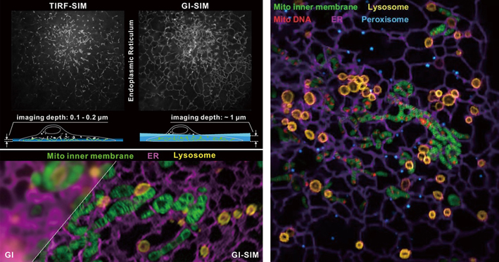
Left: GI-SIM, a new approach of microscopy, enables an in-depth and clearer visionof intracellular organelles.
Right: Multiple intracellular components and their dynamic interactions can be clearly visualized in almost real-time manner with the aid of multi-color GI-SIM. (Credit: IBP)
Seeing is the most direct way we recognize and understand our macroscopic world. This also applies when it comes to gaining insights into the microscopic world inside a cell. However, under a currently available microscopy, the intracellular world still seems too blurry.
To have a better look at what’s going on inside a cell, scientists need better imaging tools that allow noninvasive, real-time capture of what inside a cell at higher spatiotemporal resolution and lower noise background. On this regard, a new kind of microscopy, termed grazing incidence structured illumination microscopy (GI-SIM), has been developed by a joint research team, led by Prof. LI Dong from the CAS Institute of Biophysics (IBP) in Beijing and Profs. Jennifer Lippincott-Schwartz and Eric Betzig from the Howard Hughes Medical Institute in USA. This GI-SIM enables the researchers to see clearer inside a cell and gain new insights into many interesting intracellular events occurring among different organelles and cytoskeletons.
GI-SIM offers a combination of super-resolution, high-speed, multi-color imaging and low photobleaching (light-induced fading of a fluorophore) and phototoxicity (light-induced irritation or damage). These combined features make it well suited for studying intracellular dynamics. The use of GI-SIM allows the scientists to directly observe the dynamic instability of microtubules(a part of cytoskeleton) and tubular endoplasmic reticulum (a type of interconnected membrane network within cytoplasm), and to gain new insights into their mechanisms. This new imaging tool also expands our understanding of organelle-organelle hitchhiking: this scenario is now found to be mediated by membrane contact sites, which tether the hitchhiker to the vehicle organelles. Moreover, endoplasmic reticulum-mitochondria contact sites are found to promote both mitochondrial fission and fusion, with aid of this novel imaging tool.
Summed up, visualizing intracellular dynamic events in a real-time and super-resolution way opens a new window on underlying mechanisms for biological processes. “The advent of this new imaging tool may bring cell biology into a new era,” commented WANG Xiaofan, a Foreign Member of CAS and a professor from the School of Medicine, Duke University, who is not involved in this study.
Reference
Yuting Guo, Di Li, Siwei Zhang, Yanrui Yang, Jia-Jia Liu, Xinyu Wang, Chong Liu, Daniel E. Milkie, Regan P. Moore, U. Serdar Tulu, Daniel P. Kiehart, Junjie Hu, Jennifer Lippincott-Schwartz*, Eric Betzig*, Dong Li*, Visualizing Intracellular Organelle and Cytoskeletal Interactions at Nanoscale Resolution on Millisecond Timescales. Cell 175, 1430 (Published: October 25, 2018). doi: 10.1016/j.cell.2018.09.057

