

The Tan Kah Kee Young Scientist Award in Life Sciences goes to Prof. LI Hanjie from the Shenzhen Institute of Advanced Technology (SIAT) of the Chinese Academy of Sciences, recognizing his pioneering contributions to the discovery and functional characterization of a novel peripheral microglial lineage. Through high-resolution spatiotemporal mapping of early human immune development and evolutionary biology, LI’s study expands the boundaries of neuroimmunology and challenges a century-old biological dogma: proving that microglia—long thought to be exclusively confined to the brain (central nervous system)—also thrive in the peripheral nervous system, orchestrating neuronal growth and sensation across multiple species.
The feat began when LI’s team set out to map early human immune development, specifically tracking how macrophages—traditionally known for cleaning up cellular debris and responding to injury—migrate to colonize embryonic organs. While compiling this high-resolution cellular atlas, published in Cell in 2023, they stumbled upon a startling anomaly. Hidden within their massive dataset were a distinct cluster of immune cells residing far outside the brain that possessed the exact genetic, epigenetic, and developmental signatures of microglia. A major dogma in biology is that microglia exclusively live in the central nervous system (the brain and spinal cord). This study proved that wrong.
They discovered that these “microglia-like” cells thrive in the fetal skin (epidermis), testicles, and heart. They also found that, in the early fetal skin, these microglia-like cells are incredibly abundant and uniquely distributed along the back. The researchers used tissue cultures to show that these skin-based microglia actively grab onto and interact with “neural crest cells,” guiding them to turn into melanocytes (the cells that give skin its pigment).
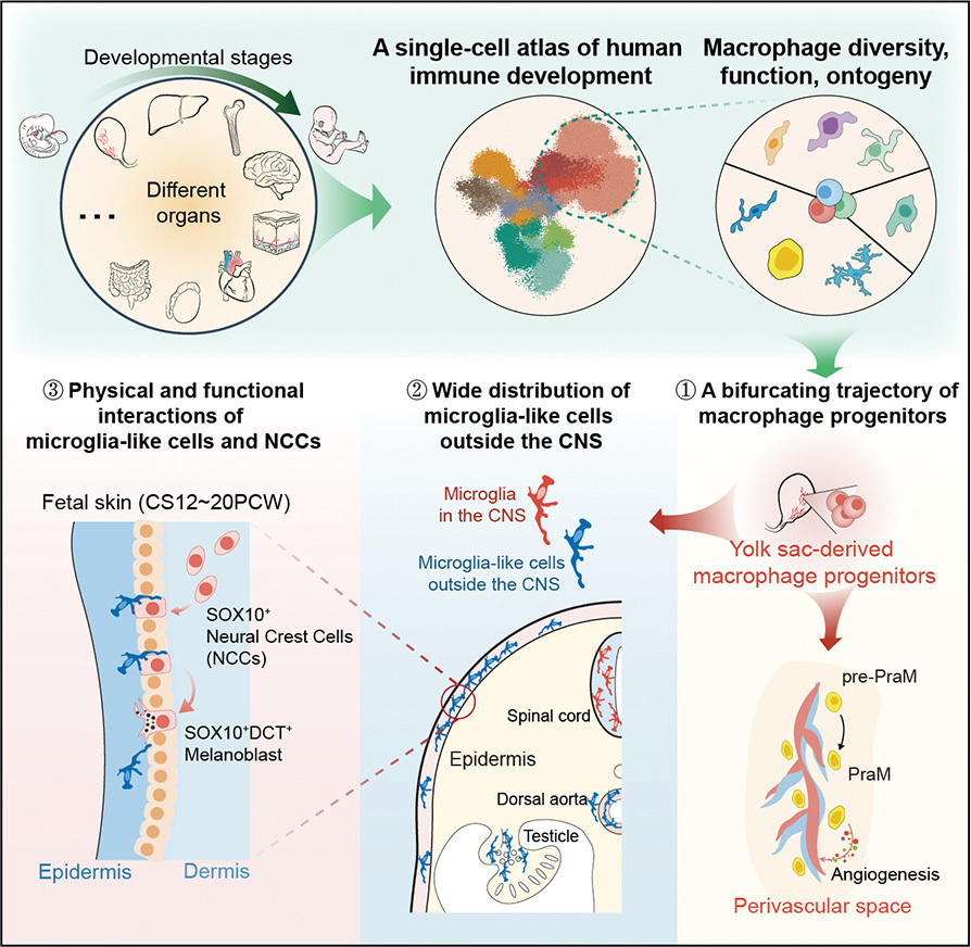
A map of early human immune development identifies specialized macrophages that help build blood vessels, alongside “microglia-like” cells that operate surprisingly far outside the central nervous system. (Graphic: Wang et al., 2023)
Notably, they also discovered a group of specialized macrophages built entirely to help construct blood vessels. The team found these proangiogenic macrophages (PraMs) residing across almost all fetal organs, but specifically hugging the outer surface of developing blood vessels. By incubating PraMs with human umbilical vein cells in a petri dish, they found that the PraM fluid actively forced the vein cells to build new blood vessel tubes.
Using computational trajectory mapping to trace the cells back in time, they discovered that specific “yolk sac-derived macrophage progenitors” undergo a fork in the road during development. One path turns these early cells into microglia and microglia-like cells, while the other path turns them into the blood-vessel-building PraMs.
Historically, microglia were believed to be strictly confined to the brain and spinal cord. After their 2023 study proved these cells also exist in human fetal skin and organs, the researchers asked the next logical question: Do these specialized immune cells also reside in the peripheral nerves that branch throughout the human body?
To answer this, in a 2025 study published in Cell, the team designed a broad phylogenetic (evolutionary) survey, collecting nervous system tissues from 24 different vertebrate species. This massive dataset included fish, amphibians, reptiles, and mammals ranging from mice to monkeys and pigs.
The researchers used advanced single-cell transcriptome sequencing (scRNA-seq) and epigenetic profiling to analyze the immune cells within the PNS. They successfully identified a unique population of PNS-resident macrophages and proved that these cells share the exact same molecular signatures, protein markers (like P2RY12 and SALL1), and developmental origins as traditional CNS microglia.
Using immunofluorescence staining, the team mapped exactly where these cells live in the physical tissue. For decades, scientists believed peripheral ganglia operated on a “duo” model consisting of only two types of cells: a neuron cell and satellite glial cells (SGCs). This study proved that the PNS microglia physically wrap themselves around the neuron’s soma (the main cell body) alongside the glial cells, establishing a brand-new Neuron-PNS Microglia-SGC trio model.
Through functional biological assays, the team discovered that these PNS microglia are absolutely required for neuronal growth. Specifically, they regulate the enlargement of the neuron’s soma and actively assist in the growth of its axons.
This is where the 24-species survey paid off, revealing the most interesting detail of the study. The team found that the presence of PNS microglia is tied directly to the physical size of the animal and its neurons, not to how closely related the species are on the evolutionary tree.
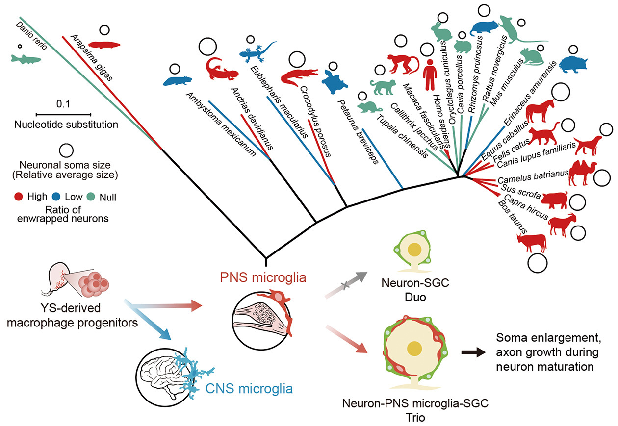
Yolk-sac macrophage progenitors develop into conserved CNS microglia and PNS microglia. These PNS microglia exist only in vertebrates with large peripheral neuronal somas, forming an essential trio with neurons and SGCs to drive soma enlargement and axon growth. (Graphic: Wu et al., 2025)
Larger animals that require massive peripheral neurons (like humans, horses, and pigs) have a high abundance of these PNS microglia to support that extreme cellular growth. Smaller animals with smaller neurons (like rodents) completely lack them. This brilliantly explains why these cells went undiscovered for so long—most global neurobiology research relies heavily on lab mice, which simply do not possess this specific cellular architecture.
The profound impact of LI’s work has been widely recognized by the global scientific community. Leading journals, including Science Immunology and Cell Research, published commentaries affirming that the human immune cell atlas and the discovery of both microglia-like cells and proangiogenic macrophages have “significantly expanded current understanding.” Nature Reviews Immunology highlighted the breakthrough in a special report titled “Microglia in the Periphery.” Furthermore, prominent experts have penned dedicated reviews praising the findings, including Marco Prinz (Member of the German National Academy of Sciences) with “Microglia out of place—mapping macrophages across the developing human body,” and Jonathan Kipnis (Member of the US National Academy of Medicine) with “Beyond the brain: microglia-like cells regulate peripheral neuronal soma size.”
The medical implications ripple far beyond the laboratory bench. If these peripheral microglia are the ultimate custodians of neural growth and sensory maintenance, what happens when they malfunction? Could their dysregulation be the hidden, long-sought culprit behind debilitating conditions like diabetic neuropathy, where peripheral nerves slowly wither, causing agonizing pain and eventual numbness? By identifying this novel cell lineage, LI’s team has effectively handed the medical world a brand-new therapeutic target—a previously invisible cellular target that might one day be turned to alleviate human suffering.
Reference
Wang, Z., Wu, Z., Wang, H., Feng, R., Wang, G., Li, M., . . . Li, H. (2023). An immune cell atlas reveals the dynamics of human macrophage specification during prenatal development. Cell, 186(20), 4454–4471.e4419. doi: 10.1016/j.cell.2023.08.019
Wu, Z., Wang, Y., Chen, W. W., Sun, H., Chen, X., Li, X., . . . Li, H. (2025). Peripheral nervous system microglia-like cells regulate neuronal soma size throughout evolution. Cell, 188(8), 2159–2174.e2115. doi: 10.1016/j.cell.2025.02.007