InBrief · 30 Dec 2025
Seeing the Whole Brain at Work
Seeing the Whole Brain at Work
When neurons fire, blood vessels nearby respond by expanding to deliver more oxygen and nutrients—a process called neurovascular coupling. This dynamic interaction is not only essential for normal brain function, but also critical for robotic limbs or computer cursors: It allows for the development of a non-invasive brain-computer interfaces to control such external devices. However, existing imaging technologies cannot simultaneously capture both neural activity and blood vessel changes across the whole cortex with sufficient speed and resolution. Researchers from the Shenzhen Institutes of Advanced Technology (SIAT) of the Chinese Academy of Sciences have now developed a microscope that overcomes these limitations.
As reported in Science Advances (doi: 10.1126/sciadv.adw5275) on July 23, the new system, called LiTA-HM (linear transducer-array-based hybrid microscope), combines multiple imaging techniques to visualize neurons and blood vessels across the whole cortex of awake mice in real time. The microscope achieves 6-micrometer resolution over a 6 mm × 5 mm field of view at 1.25 frames per second, capturing details down to individual neuron cell bodies and tiny capillaries across the entire cortex. The team successfully used LiTA-HM to study brain disease models and functional activity in awake mice. This technology provides a powerful new tool for brain research and could advance the development of non-invasive brain-computer interfaces.
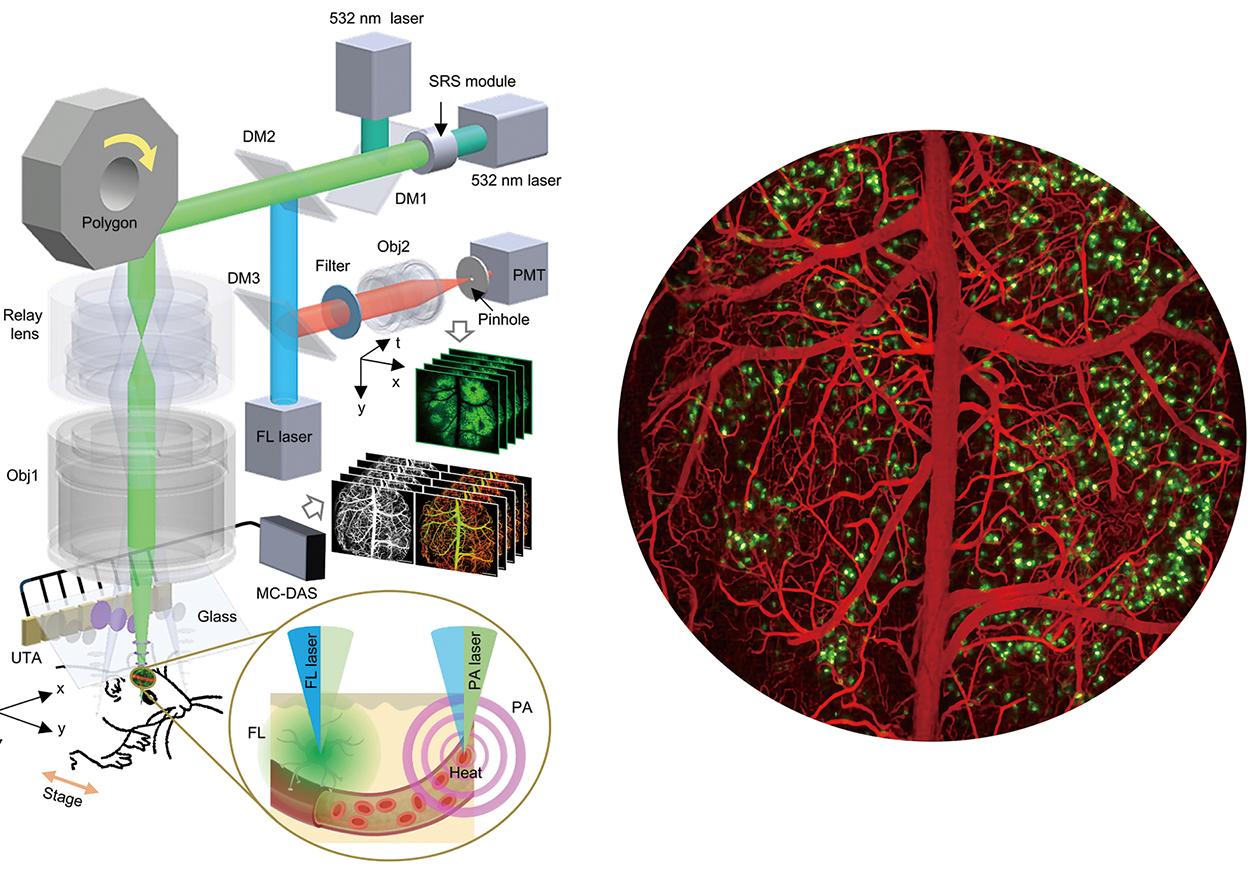
LiTA-HM schematic and imaging results. (Image by SIAT)